Document Type : Case Report
Authors
- Muhammad Priza Baktiar 1
- Manggala Pasca Wardhana 1
- Risky Vitria Prasetyo 2
- Johan Renaldo 3
- Ernawati Ernawati 1
1 Obstetrics and Gynecology Department, Dr. Soetomo General Academic Hospital, Airlangga University, Indonesia
2 Pediatric Department, Dr. Soetomo General Academic Hospital, Airlangga University, Indonesia
3 Urology Department, Dr. Soetomo General
Abstract
Introduction: Renal anomalies represent 20% of fetal congenital anomalies. Earlier diagnosis can determine proper management during pregnancy, hoping for a favorable baby outcome, mostly in developing countries.
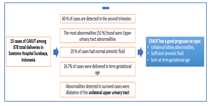
Case Report: This is a case series of Congenital Anomalies in Kidneys and Urinary Tract (CAKUT) in 2022 at Dr. Soetomo Hospital, Surabaya, Indonesia. 15 cases of CAKUT (1,7%) were reported among 878 total deliveries. All cases are referral cases from another hospital. Sixty percent of cases are detected in the second trimester. The most abnormalities found were Upper urinary tract abnormalities (53% cases). Abnormal amniotic fluid was detected in 80% of cases. 73% of cases were delivered in preterm gestational age due to spontaneous labour, premature rupture of the membrane, and fetal distress- all of these cases un-survived. Only 26.7% of cases were delivered in term gestational age, and all survived. Abnormalities detected in survived cases were dilatation of the unilateral upper urinary tract and they had normal amniotic fluid.
Conclusion: CAKUT has a good prognosis on type: unilateral kidney abnormalities, sufficient amniotic fluid, and was born at term gestational age.
Graphical Abstract
Keywords
Introduction
Fetal congenital anomalies can be anatomical or functional anomalies that arise at birth. Congenital Anomalies Kidney and Urinary Tract (CAKUT) are various migratory, structural, and functional anomalies of the kidneys, ureters, and bladder [1].
Renal anomalies represent 20% of all fetal congenital anomalies. CAKUT is the third most prevalent congenital abnormality in Europe, following cardiac anomalies and limb anomalies [2,3]. Antenatal ultrasonography (USG) plays a significant role in diagnosing CAKUT cases. The kidneys can be evaluated at 19-21 weeks of gestation, when almost all amniotic fluid consists of urine [4]. Anomalies in CAKUT can decrease the amount of amniotic fluid, leading to abnormal development of some fetal structures and a worse postnatal prognosis [3,5].
A proper diagnosis of CAKUT is essential for counseling patients on appropriate pregnancy management and planning for delivery through a multidisciplinary team approach [3]. Good antenatal care and pregnancy management have produced a term fetus and good outcome [6]. This case series aims to discover how to diagnose and manage CAKUT effectively in a developing country with limited resources to diagnose and get an excellent baby outcome.
Case series
This case series was obtained at Dr. Soetomo Hospital, Surabaya, Indonesia, in 2022. Data were obtained from hospital medical records. Almost all the cases were referral cases from peripheral hospital to the Maternal Fetal Medicine (MFM) clinic of Dr. Soetomo General Academic Hospital, the biggest referral hospital in east part of Indonesia. Most patients were referred in the gestational age range of 18-30 weeks. Following an ultrasound examination for diagnosis, there were 15 confirmed cases.
We used the Congenital Anomalies Kidney and Urinary Tract (CAKUT) classification from a study carried out in Europe by Wiesel, which categorizes CAKUT into two main categories: complex malformations and isolated or severely isolated abnormalities. Complex malformations are further divided into chromosomal, non-chromosomal, or multiple congenital anomaly syndromes, which we include in the Associated Group [7].
In 2022, Dr. Soetomo General Academic Hospital recorded 15 CAKUT cases, accounting for 1.7% of the total 878 deliveries. The distribution of the cases is presented in Table 1. There were 2 cases suspected of an association with chromosomal anomalies based on ultrasound findings; we did not do karyotype in the case series.
The management and outcomes of these case series are presented in Table 2. Almost of the cases had abnormal amniotic fluid; 11 cases detected had anhydramnios-oligohydramnios, 3 had normal amniotic fluid, and 1 had polyhydramnios. We performed amnioinfusion in 26% of cases to increase the visualization of fetal structures and improve the diagnostic.


Eight cases had dilatation of the upper urinary tract; 2 of these cases, survived after conservative management and planned deliveries in term gestational age. The postnatal evaluation showed unilateral hydronephrosis because of Vesicoureteral Reflux (VUR), and both patients managed conservatively using regular USG evaluation. In these cases, renal hydronephrosis showed improvement, and renal function tests were normal. Un-survived cases that gave birth in preterm gestational age with an indication of delivery were spontaneous in labour to hydramnios, premature rupture of the membrane (PROM), and fetal distress. All un-survived cases were not evaluated for postpartum renal abnormalities because of nonoptimal conditions, and the baby died < 24 hours after delivery.
Three cases were diagnosed with agenesis renal bilateral and also delivered preterm, with the most common indication for termination being fetal distress. Postnatal diagnosis cannot be evaluated because the baby died < 24 hours.
Two cases were suspected of chromosomal abnormalities. The first case had multiple significant congenital anomalies, i.e. fetal hydrops and bilateral renal agenesis. The baby died intrauterine at 18 weeks. The other case was skeletal dysplasia with bilateral pyelectasis; the patient delivered at 35 weeks gestation, and the baby died in < 24 hours.
One case was diagnosed with multicystic kidney (MCKD) unilateral. This case was diagnosed at 24 weeks; amniotic fluid was normal until delivery, then the patient was conservatively managed. The patient then gave birth spontaneously at 38 weeks. Postnatal diagnosis showed unilateral multicystic kidney and confirmed diagnosis using RPG showed regular renal function test.
One case was diagnosed prenatally at 31 with unilateral renal agenesis. USG found a unilateral small kidney and normal amniotic fluid. This patient was managed with conservative treatment and delivered at 39 weeks. Postnatal diagnosis revealed a normal kidney.
Discussion
Ultrasound is one of the initial screening modalities for examining fetal anomalies [8]. Kidneys can ideally be identified at 19-21 weeks of gestation [4,9]. Only 2 cases of this case series were identified in that period; most were identified in the late second trimester and third trimester. This is one of the problems in our countries and most developing countries, in which patients do not come for antenatal care as early as the pregnancy is detected. Also, the problem with the first- and the second-trimester ultrasound screening is that it is only done in some healthcare centres. It had an impact on the late diagnosis of congenital anomalies, including CAKUT.
Most of our case series showed amniotic fluid abnormality. We did amnioinfusion on anhydramnios-severe oligohydramnios cases. All types of renal abnormalities have one thing in common: the most common abnormality found is an abnormal amount of amniotic fluid. Amniotic fluid is the best indicator to assess renal function at prenatal [3]. To diagnose anomalies with inadequate amniotic fluid, we may perform amnioinfusion to improve diagnostic imaging. Amnioinfusion can increase the visualization of fetal structures from 51.0% to 77.0% and is ideally done early in the second trimester (18-20 weeks) [10].
The most common type of renal abnormality was urinary tract dilatation (Table 1), similar to studies conducted in Europe and China [7,11]. Cases of dilatation of the upper urinary tract have many etiologies, the most common being the transient type, which can heal spontaneously as much as 70% [12]. Two cases showed VUR during the postnatal evaluation. Incidence ranges from VUR 10-40% and can only diagnosed after the baby is born because it requires other examinations such as voiding cystourethrogram (VCUG). The primary management of VUR is to prevent the risk of pyelonephritis [4]. Low-grade VUR can gradually resolve with routine evaluation; high-grade VUR can be a surgical intervention that may be performed in patients with persistent reflux, renal scarring, and recurrent urinary tract infections [13].
Renal agenesis is the loss of one or both kidneys without identifiable rudimentary tissue. Its incidence in the bilateral form is 1-2/5000 for the bilateral form [9]. Renal agenesis is more common in males than females. Bilateral renal agenesis causes severe oligohydramnios and fetal or perinatal loss [14]. It can be seen from 3 cases that all cases of renal agenesis died. This can be related to the compression of the fetal chest by the diaphragm wall and the absence of amniotic fluid inhibiting lung development [9].
Individual cases of CAKUT may differ significantly in their causes. However, it has long been recognized that CAKUT often occurs in association with other congenital anomalies. Certain genetic factors, such as chromosomal abnormalities and inherited mutations, influence fetal development [15]. The most common causes of hydrops fetalis disease are a chromosomal anomaly, cystic hygroma, thorac condition, and urinary tract anomaly [16]. Skeletal system anomalies occurred in 9% of patients with CAKUT, including upper extremities and hands (4%), spine and ribs (4%), lower extremities and legs (3%), and hips and pelvis (2%) [17]. We suspected 2 cases with chromosomal anomalies due to multiple congenital malformations related to this type.
One of our survival babies was Multicystic Kidney disease (MKCD) cases. MCKD incidence is 1/1000-5000 live births, with approximately unilateral 75-80% and slightly more prevalent in males [14]. MCKD has two forms: the peripheral or central position of the more extensive cystic formation. Our case confirms that cystic is a central position after we perform a pyelogram (RPG) because we want to evaluate the function and anatomy of the kidneys. The baby's prognosis is good when found in unilateral form, and normal renal function can spontaneously resolve up to 74% of patients at the age of two years [14].
Renal dysplasia is variable in size, but most are smaller than usual. Incidence is 1/1000 newborns for the unilateral form [9]. The male-to-female ratio for unilateral is 1.9: 1. Unilateral renal agenesis is usually asymptomatic and can be detected incidentally during routine ultrasound examination [18].
The gender ratio is male and female at 3:1. The male gender dominates these cases but also has a high spontaneous cure rate [19]. Gender-specific mechanisms still require further research and are currently associated with environmental factors, renal transcriptomes, and gene expression and pathways [1].
Baby outcomes were 11 cases of death, with 10 cases of dying < 24 hours (Table 2). The un-survived babies were delivered at preterm gestational age, had abnormal amniotic fluid and some of them had multiple congenital anomalies. Preterm gestational age delivery is another factor in the prognosis of a baby. Preterm is related to 27% of infant mortality [20]. Amniotic fluid showed renal function and maternal-fetal circulation. Inadequate amniotic fluid may result in the abnormal development of several fetal structures, one of which is lung hypoplasia [3]. Normal amniotic fluid may show the functioning of unilateral or bilateral renal of the babies, which shows the acquired anomalies, only affecting one kidney and no other anomalies.
To our knowledge, this is the first case series on CAKUT in the Indonesian population, showing the babies' diagnosis and management until delivery. Some publications reported from develops country data [1,2]. However, this case was a series of retrospective cases, which impacted incomplete data in some cases, and some patients could not be contacted to confirm diagnoses.
Conclusion
Proper diagnosis and appropriate treatment of congenital anomalies of the kidney and urinary tract (CAKUT) have been shown to influence fetal outcomes greatly. The occurrences of abnormalities, adequate amniotic fluid, unilateral kidney malformations, and delivery at term gestational age contribute to a more favourable outcome. Fetus with unilateral renal problems, sufficient amniotic fluid levels, and being delivered at full term gestational age have a better prognosis.
Orcid:
Muhammad Priza Baktiar: https://orcid.org/0009-0003-0501-7872
Manggala Pasca Wardhana: https://www.orcid.org/0000-0001-8013-4639
Risky Vitria Prasetyo: https://www.orcid.org/0000-0002-1708-5384
Johan Renaldo: https://www.orcid.org/0000-0001-6001-498x
Ernawati Ernawati*: https://www.orcid.org/0000-0002-9344-3606
-----------------------------------------------------------------------
How to cite this article: Muhammad Priza Baktiar , Manggala Pasca Wardhana , Risky Vitria Prasetyo, Johan Renaldo, Ernawati Ernawati* ,Problem in diagnosis and management of congenital anomalies kidney and urinary tract (cakut) in developing countries: a case series. Journal of Medicinal and Pharmaceutical Chemistry Research, 2024, 6(4), 474-480. Link: http://jmpcr.samipubco.com/article_186356.html
-----------------------------------------------------------------------
Copyright © 2024 by SPC (Sami Publishing Company) + is an open access article distributed under the Creative Commons Attribution License(CC BY) license (https://creativecommons.org/licenses/by/4.0/), which permits unrestricted use, distribution, and reproduction in any medium, provided the original work is properly cited.


