Document Type : Original Research Article
Authors
- Ingrid Melinda Prasetyanto 1
- R.A. Meisy Andriana 2
- I Putu Alit Pawana 3
- Damayanti Tinduh 3
- Hermina Novida 4
- Budi Utomo 5
1 Resident of Department of Physical Medicine and Rehabilitation, Faculty of Medicine, Airlangga University, Surabaya, Indonesia, 60264
2 Neuromuscular Division, Department of Physical Medicine and Rehabilitation, Faculty of Medicine, Airlangga University, Surabaya, Indonesia, 60264
3 Sport Injury Division, Department of Physical Medicine and Rehabilitation, Faculty of Medicine, Airlangga University, Surabaya, Indonesia, 60264
4 Endocrine and Metabolic Division, Department of Internal Medicine, Faculty of Medicine, Airlangga University, Surabaya, Indonesia, 60264
5 Epidemiology and Biostatistic Division, Department of Public Health and Preventive Medicine, Faculty of Medicine, Airlangga University, Surabaya, Indonesia
Abstract
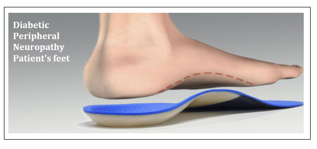
Background: Treatment that can be done to prevent ulceration of the diabetic foot is to reduce areas of high foot pressure. One strategy that is being developed is the use of insoles. Scientific evidence regarding the use of insoles is still sparse, but the benefits of soft insoles can improve function, distribute foot pressure, and protect/prevent ulceration.
Objective: This study aims to compare and analyse peak plantar pressure using PEDAR-X® in Type 2 Diabetes Mellitus patient with diabetic neuropathy after administration of both insoles.
Method: Pre-experimental study with 2 groups pre- and post-intervention providing custom-made and prefabricated insoles. A total of 32 subjects with diabetic neuropathy were divided into 2 groups, and then the plantar pressure was measured at the beginning of the examination, and immediately after administration of the insole in each study group.
Results: There was a reduction in peak plantar pressure using the PEDAR-X® device in type 2 Diabetes Mellitus patient with diabetic neuropathy after administering custom made and prefabricated insoles. The effect of providing custom-made insoles is more beneficial than prefabricated insoles, seen from the more uniform decrease in custom-made insoles, although both still provide a significant decrease effect.
Conclusion: Significant reduction in average plantar pressure in the heel, forefoot, and overall areas in both insoles. This supports the recommendation for giving soft insoles to patients with diabetic neuropathy.
Graphical Abstract
Keywords
Main Subjects
Introduction
One of the complications of diabetes mellitus that causes high mortality and morbidity is diabetic foot [1]. Diabetic foot may lead to diabetic ulcer and lower extremity amputation [2]. The underlying mechanisms of diabetic foot in type 2 diabetes mellitus are peripheral nerve damage, endothelial injury and vascular dysfunction [3]. The average duration of diabetes is longer in patients with more severe neuropathy and risk of ulcer formation increases over time [4]. Increased plantar pressure and peripheral sensorimotor neuropathy with loss of protective sensation in the feet are the most important factors in diabetic foot ulceration [5]. Approximately 94% of diabetic ulcers occur in areas of increased plantar pressure. Successful healing and prevention of foot ulcers depends on reducing high foot pressure [6]. Inadequate foot sensation can be due to neuropathy, which results in the patient's feedback (proprioception and pain) in assessing independent foot pressure reduction, which is indicated by changing gait patterns, resting, or removing shoes, to be reduced as well [7]. Peripheral Neuropathy may also lead to poor control of muscle strength that increases the risk of fall [8].
Plantar pressure distribution measurements are used to determine the pressure between the plantar surface of the foot and the sole of the shoe. There are 2 types of foot plantar pressure measurements, platform system and in-shoe pressure sensors [9]. In-shoe pressure sensors have paved the way towards better systems measuring efficiency, flexibility, mobility, and cost reduction. These in-shoe sensors are flexible and can be installed inside the shoe in such a way that measurements can reflect the surface of the foot and shoe. The PEDAR-X® system is a portable examination and can enable a wider range of studies [10]. Using PEDAR-X® for just 8-12 steps per foot can record the patient's plantar pressure profile. Measuring plantar pressure distribution using PEDAR-X® can also measure peak pressure, maximum force, pressure time integral, and contact area and is a non-invasive examination that can detect ulcer-prone areas on the soles of the feet of type 2 DM patients that are not clinically visible [11].
There are various strategies to reduce plantar pressure including casting, corrective orthotic devices such as insoles, rockers, and therapeutic shoes [12]. The main principles of providing footwear and orthoses are for off-loading, ulceration protection, and ensuring a person uses footwear that fits properly. Footwear that can be used can include modified footwear, therapeutic shoes, and orthoses, depending on the position of the ulceration. Not all diabetes patients require therapeutic footwear, but most will benefit from some type of insole or orthosis to improve function, redistribute foot pressure and/or provide additional cushioning/protection [13]. Research regarding the choice of material, shape, and features of insoles for diabetic feet has been widely studied [14], but due to the large heterogeneity of these studies, it is difficult to make it the main choice for diabetic foot patient [15]. Insoles available on the market vary in terms of material, thickness, additional features (metatarsal pads, arch, heel pads), comfort, and price. Research by Haris et al. (2021) shows that the ethylene-vinyl acetate (EVA) material in insoles is most often used, and can reduce 37% peak plantar pressure in subjects with diabetes mellitus [16]. The use of arch support in insoles was also recommended by Collings et al. (2019) because through their study on samples with diabetes mellitus, there was a decrease in peak pressure of 37 kPa when compared to flat insoles [17]. A flat and soft insole was found to reduce peak pressure and maximize the contact area on the foot surface. Custom-made insoles are said to be more effective than soft insoles, especially if the insole is contoured in the arch support area [18]. This study compares insoles available in the marketplace with insoles made in the orthosis prosthesis workshop at Dr. Soetomo Hospital, Surabaya, Indonesia.
Materials and methods
This research is a pre-post test study without a control group with a quasi-experimental study design. The study was carried out at the Medical Rehabilitation Installation at Dr. Soetomo Surabaya starting January-May 2023. This study received a certificate of ethical suitability from the Health Research Ethics Committee of Dr. Soetomo General Hospital Surabaya with no. 0619/KEPK/III/2023. The research subjects were controlled type 2 DM patient who visited the Endocrine Polyclinic and/or Medical Rehabilitation Polyclinic at Dr. Soetomo General Hospital. Inclusion criteria in this study include: 1) Controlled type 2 DM patients who meet the DM diagnosis criteria according to PERKENI 2019, 2) presence of diabetic neuropathy that meets the Michigan Neuropathy Screening Instrument (MNSI) criteria, 3) age 18-59 years, 4 ) normal cognitive function, 5) able to walk without a walking aid or prosthesis, 6) corrected vision, and 7) willing to participate in this research voluntarily by signing a consent form to become a research subject (informed consent). Exclusion criteria in this study include: 1) Ulcers or open wounds on one or both feet, and Charcot foot, 2) neuromusculoskeletal disease in the lower limbs that interferes with ambulation function, 3) systolic blood pressure > 160 and/or diastolic > 90 mmHg at rest, pulse > 120x/minute, body temperature > 37.5 oC, and SpO2 < 95%, 4) hypoglycemia (blood glucose < 70 mg/dL) or characterized by symptoms including shaking, weakness, excessive sweating normal, nervous, anxious, tingling in the mouth and fingers, and feeling hungry, 5) hyperglycemia (blood glucose ≥ 250 mmHg) or characterized by symptoms of polyuria, fatigue, weakness, increased thirst, and acetone breath, 6) cardiorespiratory complaints that affecting physical performance during data collection (NYHA class 3-4 heart failure, acute heart attack, unstable angina, uncontrolled arrhythmia, aneurysm, severe aortic stenosis, acute endocarditis/pericarditis, persistent asthma, and acute or severe respiratory failure). Criteria for dropping out of the test include if the subject states that he has withdrawn or died.
Subjects were given information about the aims and objectives of the study. Subject are asked to sign a research consent form (informed consent) if they are willing to become research subjects. Data collection on subject characteristics (name and age), subjective examination (anamnesis) and physical examination, as well as other examinations necessary to determine inclusion and exclusion criteria. Subjects then underwent the Michigan Neuropathy Screening Instrument (MNSI) examination. Subjects were dressed in standard socks and shoes according to their foot size. The subject is worn with the PEDAR-X® device and asked to walk at a speed that is comfortable for the subject, at least 20 steps. The subject will be helped to maintain the walking speed using a metronome. Subjects continued with an in-sole plantar pressure measurement system examination using the PEDAR-X® tool, to measure Peak Plantar Pressure (PPP) using the masking method. The examination begins with an initial examination of the plantar pressure distribution. Four trials were carried out, each with 10 steps. The areas of highest plantar pressure on the left and right feet were determined by the researchers, which were considered to be at risk for foot ulceration and were targeted for off-loading during learning sessions. Insoles were given to both groups, divided into 2 groups, those using custom-made and prefabricated insoles. PPP under the risk area as well as PPP under all other areas of both feet were measured at baseline (T0) and at the end of the study period after administration of insoles (T1).
Results
The total subjects were 32 subjects, who were divided into group 1 who received prefabricated insoles (n=16 subjects / 32 feet) and group 2 who received custom made insoles (n=16 subjects / 32 feet). In group 1, the sample size was 12 men (18.8%) and 20 women (15.6%) while in group 2, the sample size was 10 men (15.6%) and women as many as 22 people (34.4%) (p = 0.599). The mean age of patients in group 1 was 50.50 ± 7.11 years with an age range between 34-59 years, while in group 2 it was 48.18±1.63 years with an age range between 25-59 years (p = 0.071). The mean body weight of group 1 was 64.25±13.33 kg with a weight range between 46-96 kg, while group 2 was 61.46 ± 10.55 kg with a body weight range of 43.50-82 kg (p = 0.273). The mean height of group 1 was 154.37±8.73 cm with a height range of 140-171 cm, while group 2 was 153.68±6.86 cm with a height range of 142-169 cm (p = 0.246). The mean BMI of group 1 was 27.06 ± 5.71 kg/m2 with a BMI range of 19.40-37.80 kg/m2, while group 2 was 26.05±4.24 kg/m2 with a BMI range of 19.60-33.20 kg/m2 (p = 0.01). The mean HbA1c of group 1 was 7.09 ± 1.78% with an HbA1c range of 5.0-11.1%, while group 2 was 8.65±2.66% with an HbA1c range of 5.3-15.9% (p = 0.068). The mean diabetes onset of group 1 was 7.50±6.96 years with a diabetes onset range of 0.33 - 23 years, while group 2 was 5.64±3.74 years with a diabetes onset range of 0.75-13 years (p = 0.002). The average walking speed of group 1 was 91.31±3.08 m/s with a walking speed range of 87-98 m/s, while group 2 was 92.12 ± 3.96 m/s with a walking speed range of 85-100 m/s. s (p = 0.202). No significant difference in peak plantar pressure in both groups at the beginning of the study. The characteristic of the subjects is presented in Table 1.
There were no significant differences of peak plantar pressure in both group at the end of study (Table 2), but there was a significant decrease of mean peak plantar pressure value at mask 1 (heel), mask 3 (forefoot), and total peak plantar pressure after being given the prefabricated insole in both groups (Table 3). There was also significant delta of peak plantar pressure at mask 1 (heel), mask 3 (forefoot), and total peak plantar pressure in both groups (Table 4). The effect size of giving prefabricated insoles and custom size insoles was calculated using Cohen's d and shown in Table 5.
Discussion
Changes in peak plantar pressure values in type 2 DM patients are important risk factors for diabetic ulcers. Increase in pressure is proportional to an increase in the possibility of ulcers [11,19]. A study from the pressure of 60 N/cm2 was the upper threshold for the occurrence of ulcers in diabetes mellitus [20]. Hazari et al. (2019) set a cutoff point of 335 kPa (peak plantar pressure) as the risk threshold for ulceration in the forefoot area [21]. Several other studies apply pressure thresholds of up to 600 kPa [5,22], 1000 kPa, and 1230 kPa [23]. Higher plantar pressure accompanied by sensory and motor neuropathy can be a potential risk for foot ulceration [21]. Higher plantar pressure can be significantly associated with deformity and soft tissue changes in the foot. Diabetic neuropathy patients can develop ulcerations even at normal plantar pressure values [24,25]. The findings from this study may help strengthen the importance of plantar pressure threshold values with future experimental studies. This factor becomes more important when the biomechanical condition of the ulcerated patient is considered.
The heel segment determines the plantar pressure in the heel-strike phase, while the midfoot and forefoot segments determine the plantar pressure in the mid-strike phase. The toes determine the plantar pressure in the push-off phase. This study shows that the highest mean peak plantar pressure is located on the forefoot (195.22±39.10 mmHg in the prefabricated group and 162.26±46.84 mmHg in the custom made group) and is followed by pressure on the heel. This is in accordance with the results of several studies, such as an increase in diabetic foot pressure patterns in the forefoot/metatarsal area in a study by Lord and Hosein [26]. A number of factors have been described as increasing foot pressure in the forefoot area, such as the thickness of the tissue/fat pad [27], and the protrusion of the metatarsal bones [28]. Compared with non-diabetic subjects, patients with diabetic neuropathy show a decrease in active ankle ROM and dynamic ankle flexion in the heel-strike phase and a decrease in ROM (flexion-extension) amplitude resulting in increased pressure in the heel area [29].
Diabetic peripheral nerve damage and vascular disease cause gradual changes in pressure on the metatarsals which initially decreases because the load is handled by the hallux, then increases because then excess pressure is diverted from the hallux (relatively insensitive, flexible, and unable to withstand too large gradual loads) [27]. Patients with neurovascular disorders with impaired protective sensation are unable to perceive high pressure and adjust their weight-bearing activities, which causes a relationship between the most common location of ulcers and the distribution of plantar pressure [30].
Changes in peak plantar pressure values were also found after administration of both insoles. Characteristics of peak plantar pressure values before therapy for all masks showed no significant differences between the means of the two groups with a p-value > 0.05. In the prefabricated insole and custom made insole groups, significant improvements were obtained in mask 1 (heel), mask 3 (forefoot), and total, with a p-value <0.05. These results are in line with other research by Tang et al. (2014) who conducted research on patients with diabetic neuropathy in Switzerland, by comparing 3 types of insoles with different thicknesses. Decrease in peak plantar pressure values was found in the big toe, forefoot, midfoot and heel with the most significant changes found in the heel. A thick insole can hold the heel cushion under the protrusion of the calcaneus bone which in turn has a pressure reducing effect [31]. Shi et al. (2022) compared 5 types of insoles in 22 patients with diabetic neuropathy, explaining what is in accordance with this study where the use of insoles can significantly reduce peak plantar pressure values in the forefoot and heel / rearfoot areas [32].
This study has limitations. First, this study measure peak plantar pressure immediately after insole administration and requires long-term periodic measurements. Future research needed to consider periodic and long-term evaluations to assess the possibility of foot ulcers/re-ulceration in diabetes patient, related to the durability of the used materials, length of daily use, and development/addition of insole features such as arch support, metatarsal pads, or contoured insoles, or by comparing other offloading therapies, as well as comparing the effects of using insoles with standard or currently used therapies. Bias factors that are still difficult for researchers to control are physical activity, daily intake, adherence to treatment and the habits and conditions of each subject. These four factors can influence plantar pressure and the distribution of pressure. The subject's habits include walking patterns, use of inappropriate footwear, thickness of pads in the foot fat, and foot care. Further research is needed to monitor these factors as controlled bias variables.
Conclusion
The use of both types of insoles in diabetic neuropathy patients provides an immediate effect of significantly reducing peak plantar pressure in the total area, heel, and big toe. There was no difference in peak plantar pressure in the two groups after using the two types of insoles. However, the use of custom made insoles provided a more significant and even reduction in peak plantar pressure compared to prefabricated insoles.
ORCID
Ingrid Melinda Prasetyanto*: https://www.orcid.org/0009-0006-4631-7136
R.A. Meisy Andriana: https://www.orcid.org/0000-0003-3299-0179
I Putu Alit Pawana: https://www.orcid.org/0000-0002-6775-964X
Damayanti Tinduh: https://www.orcid.org/0000-0001-6604-8152
Hermina Novida: https://www.orcid.org/0000-0001-5899-1589
Budi Utomo: https://www.orcid.org/0000-0001-6060-9190
---------------------------------------------------------------------------------
How to cite this article: Ingrid Melinda Prasetyanto*, R.A. Meisy Andriana, I Putu Alit Pawana, Damayanti Tinduh, Hermina Novida, Budi Utomo , Insoles reduce peak plantar pressure in diabetic peripheral neuropathy. Journal of Medicinal and Pharmaceutical Chemistry Research, 2024, 6(5), 571-580. Link: http://jmpcr.samipubco.com/article_187421.html
---------------------------------------------------------------------------------
Copyright © 2024 by SPC (Sami Publishing Company) + is an open access article distributed under the Creative Commons Attribution License(CC BY) license (https://creativecommons.org/licenses/by/4.0/), which permits unrestricted use, distribution, and reproduction in any medium, provided the original work is properly cited.


.png)
.png)
.png)
